A photonic crystal is an optical nanostructure in which the refractive index changes periodically. This affects the propagation of light in the same way that the structure of natural crystals gives rise to X-ray diffraction and that the atomic lattices of semiconductors affect their conductivity of electrons. Photonic crystals occur in nature in the form of structural coloration and animal reflectors, and, as artificially produced, promise to be useful in a range of applications.
The term biophotonics denotes a combination of biology and photonics, with photonics being the science and technology of generation, manipulation, and detection of photons, quantum units of light. Photonics is related to electronics and photons. Photons play a central role in information technologies, such as fiber optics, the way electrons do in electronics.

Silicon photonics is the study and application of photonic systems which use silicon as an optical medium. The silicon is usually patterned with sub-micrometre precision, into microphotonic components. These operate in the infrared, most commonly at the 1.55 micrometre wavelength used by most fiber optic telecommunication systems. The silicon typically lies on top of a layer of silica in what is known as silicon on insulator (SOI).
An ultrashort pulse laser is a laser that emits ultrashort pulses of light, generally of the order of femtoseconds to one picosecond. They are also known as ultrafast lasers owing to the speed at which pulses "turn on" and "off"—not to be confused with the speed at which light propagates, which is determined by the properties of the medium, particularly its index of refraction, and can vary as a function of field intensity and wavelength.
A spaser or plasmonic laser is a type of laser which aims to confine light at a subwavelength scale far below Rayleigh's diffraction limit of light, by storing some of the light energy in electron oscillations called surface plasmon polaritons. The phenomenon was first described by David J. Bergman and Mark Stockman in 2003. The word spaser is an acronym for "surface plasmon amplification by stimulated emission of radiation". The first such devices were announced in 2009 by three groups: a 44-nanometer-diameter nanoparticle with a gold core surrounded by a dyed silica gain medium created by researchers from Purdue, Norfolk State and Cornell universities, a nanowire on a silver screen by a Berkeley group, and a semiconductor layer of 90 nm surrounded by silver pumped electrically by groups at the Eindhoven University of Technology and at Arizona State University. While the Purdue-Norfolk State-Cornell team demonstrated the confined plasmonic mode, the Berkeley team and the Eindhoven-Arizona State team demonstrated lasing in the so-called plasmonic gap mode. In 2018, a team from Northwestern University demonstrated a tunable nanolaser that can preserve its high mode quality by exploiting hybrid quadrupole plasmons as an optical feedback mechanism.
A random laser (RL) is a laser in which optical feedback is provided by scattering particles. As in conventional lasers, a gain medium is required for optical amplification. However, in contrast to Fabry–Pérot cavities and distributed feedback lasers, neither reflective surfaces nor distributed periodic structures are used in RLs, as light is confined in an active region by diffusive elements that either may or may not be spatially distributed inside the gain medium.
Time Stretch Microscopy also known as Serial time-encoded amplified imaging/microscopy or stretched time-encoded amplified imaging/microscopy' (STEAM) is a fast real-time optical imaging method that provides MHz frame rate, ~100 ps shutter speed, and ~30 dB optical image gain. Based on the Photonic Time Stretch technique, STEAM holds world records for shutter speed and frame rate in continuous real-time imaging. STEAM employs the Photonic Time Stretch with internal Raman amplification to realize optical image amplification to circumvent the fundamental trade-off between sensitivity and speed that affects virtually all optical imaging and sensing systems. This method uses a single-pixel photodetector, eliminating the need for the detector array and readout time limitations. Avoiding this problem and featuring the optical image amplification for dramatic improvement in sensitivity at high image acquisition rates, STEAM's shutter speed is at least 1000 times faster than the state - of - the - art CCD and CMOS cameras. Its frame rate is 1000 times faster than fastest CCD cameras and 10 - 100 times faster than fastest CMOS cameras.
Demetri Psaltis is a Greek-American electrical engineer who was the Dean of the School of Engineering at École Polytechnique Fédérale de Lausanne from 2007 to 2017. He is a professor in Bioengineering and Director of the Optics Laboratory of the EPFL. He is one of the founders of the term and the field of optofluidics. He is also well known for his past work in holography, especially with regards to optical computing, holographic data storage, and neural networks. He is an author of over 1100 publications, contributed more than 20 book chapters, invented more than 50 patents, and currently has a h-index of 98.

Live-cell imaging is the study of living cells using time-lapse microscopy. It is used by scientists to obtain a better understanding of biological function through the study of cellular dynamics. Live-cell imaging was pioneered in the first decade of the 21st century. One of the first time-lapse microcinematographic films of cells ever made was made by Julius Ries, showing the fertilization and development of the sea urchin egg. Since then, several microscopy methods have been developed to study living cells in greater detail with less effort. A newer type of imaging using quantum dots have been used, as they are shown to be more stable. The development of holotomographic microscopy has disregarded phototoxicity and other staining-derived disadvantages by implementing digital staining based on cells’ refractive index.

Plasmonics or nanoplasmonics refers to the generation, detection, and manipulation of signals at optical frequencies along metal-dielectric interfaces in the nanometer scale. Inspired by photonics, plasmonics follows the trend of miniaturizing optical devices, and finds applications in sensing, microscopy, optical communications, and bio-photonics.
Integrated quantum photonics, uses photonic integrated circuits to control photonic quantum states for applications in quantum technologies. As such, integrated quantum photonics provides a promising approach to the miniaturisation and scaling up of optical quantum circuits. The major application of integrated quantum photonics is Quantum technology:, for example quantum computing, quantum communication, quantum simulation, quantum walks and quantum metrology.

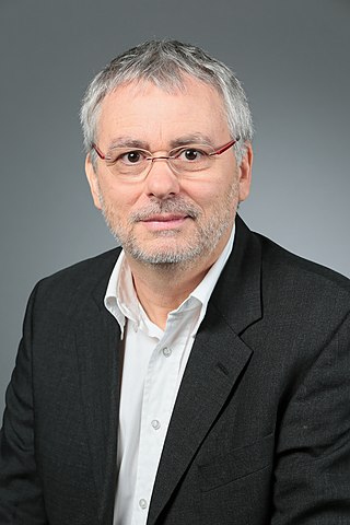
Juerg Leuthold is a full professor at ETH Zurich, Switzerland.
Kerr frequency combs are optical frequency combs which are generated from a continuous wave pump laser by the Kerr nonlinearity. This coherent conversion of the pump laser to a frequency comb takes place inside an optical resonator which is typically of micrometer to millimeter in size and is therefore termed a microresonator. The coherent generation of the frequency comb from a continuous wave laser with the optical nonlinearity as a gain sets Kerr frequency combs apart from today’s most common optical frequency combs. These frequency combs are generated by mode-locked lasers where the dominating gain stems from a conventional laser gain medium, which is pumped incoherently. Because Kerr frequency combs only rely on the nonlinear properties of the medium inside the microresonator and do not require a broadband laser gain medium, broad Kerr frequency combs can in principle be generated around any pump frequency.

Photoacoustic microscopy is an imaging method based on the photoacoustic effect and is a subset of photoacoustic tomography. Photoacoustic microscopy takes advantage of the local temperature rise that occurs as a result of light absorption in tissue. Using a nanosecond pulsed laser beam, tissues undergo thermoelastic expansion, resulting in the release of a wide-band acoustic wave that can be detected using a high-frequency ultrasound transducer. Since ultrasonic scattering in tissue is weaker than optical scattering, photoacoustic microscopy is capable of achieving high-resolution images at greater depths than conventional microscopy methods. Furthermore, photoacoustic microscopy is especially useful in the field of biomedical imaging due to its scalability. By adjusting the optical and acoustic foci, lateral resolution may be optimized for the desired imaging depth.

Stephen A. Boppart is a principal investigator at the Beckman Institute for Advanced Science and Technology at the University of Illinois at Urbana-Champaign, where he holds an Abel Bliss Professorship in engineering. He is a faculty member in the departments of electrical and computer engineering, bioengineering, and internal medicine. His research focus is biophotonics, where he has pioneered new optical imaging technologies in the fields of optical coherence tomography, multi-photon microscopy, and computational imaging.
Elizabeth M. C. Hillman is a British-born academic who is Professor of Biomedical Engineering and Radiology at Columbia University. She was awarded the 2011 Adolph Lomb Medal from The Optical Society and the 2018 SPIE Biophotonics Technology Innovator Award.
Three-photon microscopy (3PEF) is a high-resolution fluorescence microscopy based on nonlinear excitation effect. Different from two-photon excitation microscopy, it uses three exciting photons. It typically uses 1300 nm or longer wavelength lasers to excite the fluorescent dyes with three simultaneously absorbed photons. The fluorescent dyes then emit one photon whose energy is three times the energy of each incident photon. Compared to two-photon microscopy, three-photon microscopy reduces the fluorescence away from the focal plane by , which is much faster than that of two-photon microscopy by . In addition, three-photon microscopy employs near-infrared light with less tissue scattering effect. This causes three-photon microscopy to have higher resolution than conventional microscopy.

Choi Wonshik is an optical physicist researching deep-tissue imaging and imaging through scattering media. He is a full professor in the Department of Physics of Korea University where he serves as the associate director at the IBS Center for Molecular Spectroscopy and Dynamics. Inside the Center, he leads the Super-depth Imaging Lab. He has been cited more than 4,000 times and has an h-index of 32. He is a fellow of The Optical Society and the Korean Academy of Science and Technology.

Luc Thévenaz is a Swiss physicist who specializes in fibre optics. He is a professor of physics at EPFL and the head of the Group for Fibre Optics School of Engineering.
Gabriel Popescu was a Romanian-American optical engineer, who was the William L. Everitt Distinguished Professor in Electrical and Computer Engineering at University of Illinois Urbana-Champaign. He was best known for his work on biomedical optics and quantitative phase-contrast microscopy.


